Abstract
Objectives: To investigate regional differences in grey matter volume associated with the practice of Sahaja Yoga Meditation.
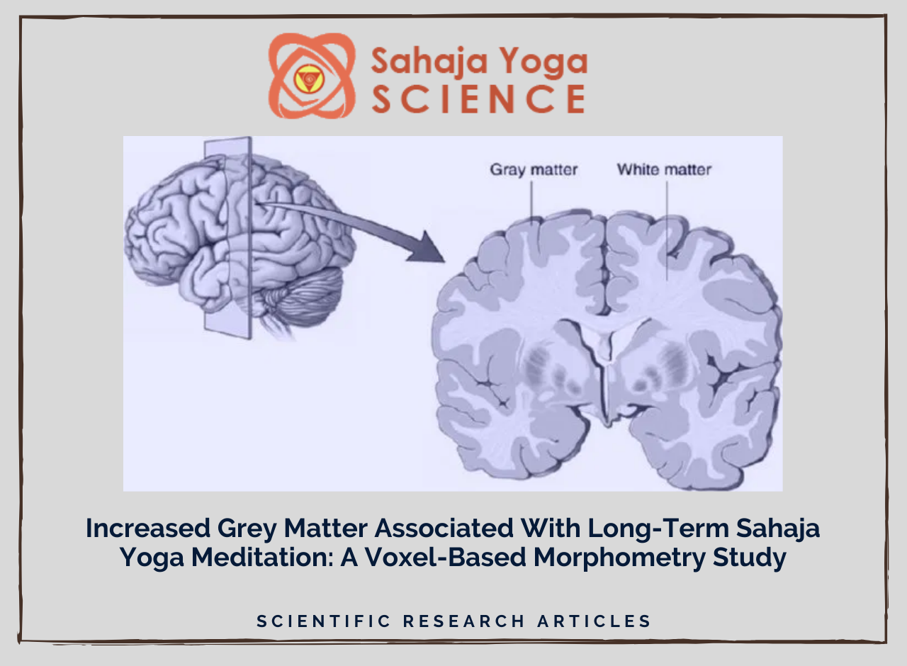
Design: Twenty three experienced practitioners of Sahaja Yoga Meditation and twenty three non-meditators matched on age, gender and education level, were scanned using structural Magnetic Resonance Imaging and their grey matter volume were compared using Voxel-Based Morphometry.
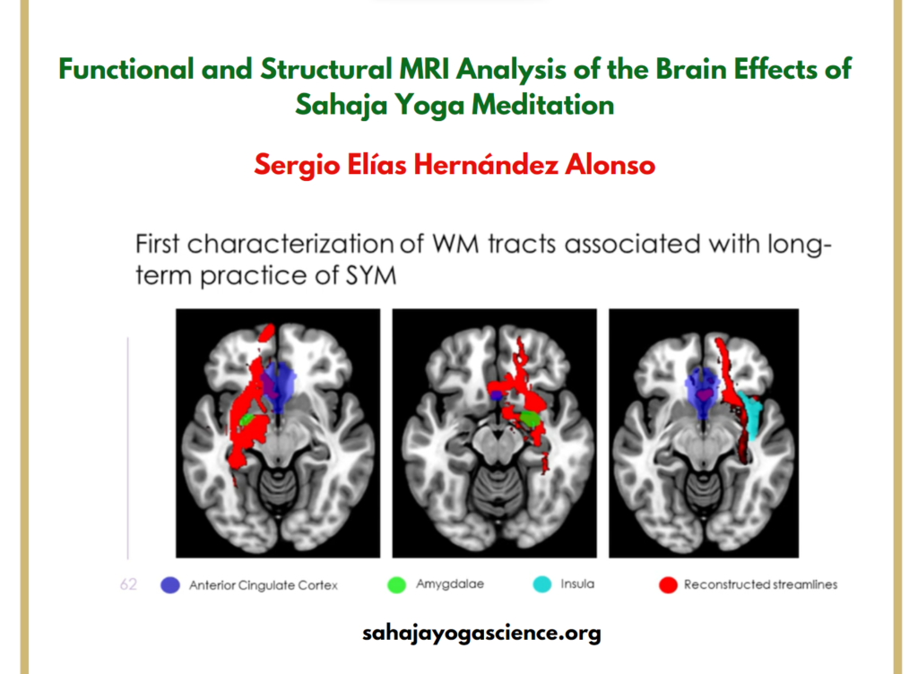
Results: Grey matter volume was larger in meditators relative to non-meditators across the whole brain. In addition, grey matter volume was larger in several predominantly right hemispheric regions: in insula, ventromedial orbitofrontal cortex, inferior temporal and parietal cortices as well as in left ventrolateral prefrontal cortex and left insula. No areas with larger grey matter volume were found in non-meditators relative to meditators.
Conclusions: The study shows that long-term practice of Sahaja Yoga Meditation is associated with larger grey matter volume overall, and with regional enlargement in several right hemispheric cortical and subcortical brain regions that are associated with sustained attention, self-control, compassion and interoceptive perception. The increased grey matter volume in these attention and self-control mediating regions suggests use-dependent enlargement with regular practice of this meditation.
Sergio Elías Hernández 1, José Suero 2, Alfonso Barros 3, José Luis González-Mora 4, Katya Rubia 5
Figures
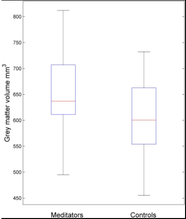
Fig 1. Box plot of grey matter volumes across the whole brain of meditators and controls. The central box represents the value from the lower to upper quartile (25th to 75th percentile). The middle line represents the median. The vertical line extends from the minimum to the maximum value.
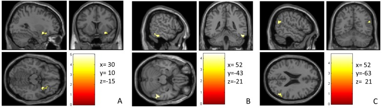
Fig 2. Areas where meditators exhibited larger GMV than non-meditators. (A) right insula/vmOFC, (B) right inferior temporal gyrus and (C) right angular gyrus. The color intensity represents T-statistic values at the voxel level.
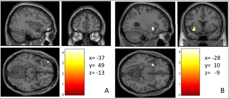
Fig 3. A priori hypothesised regions that showed larger GMV in meditators relative to controls at a more lenient threshold. (A) left VLPFC (B) left anterior insula. The color intensity represents T-statistic values at the voxel level.
Affiliations
- 1Department of Ingeniería Industrial, Universidad de La Laguna, Tenerife, Spain.
- 2Centro de Salud Jazmín, Sermas, Madrid, Spain.
- 3Department of Psychology, Universitat Jaume I, Castellón, Spain.
- 4Department of Fisiología, Universidad de La Laguna, Tenerife, Spain.
- 5Institute of Psychiatry, Psychology and Neuroscience, King’s College London, London, United Kingdom.
- PMID: 26938433
- PMCID: PMC4777419
- DOI: 10.1371/journal.pone.0150757
Source – https://pubmed.ncbi.nlm.nih.gov/26938433/#affiliation-3